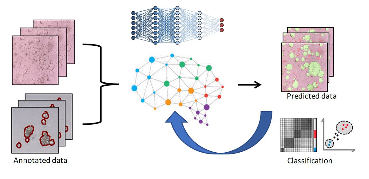
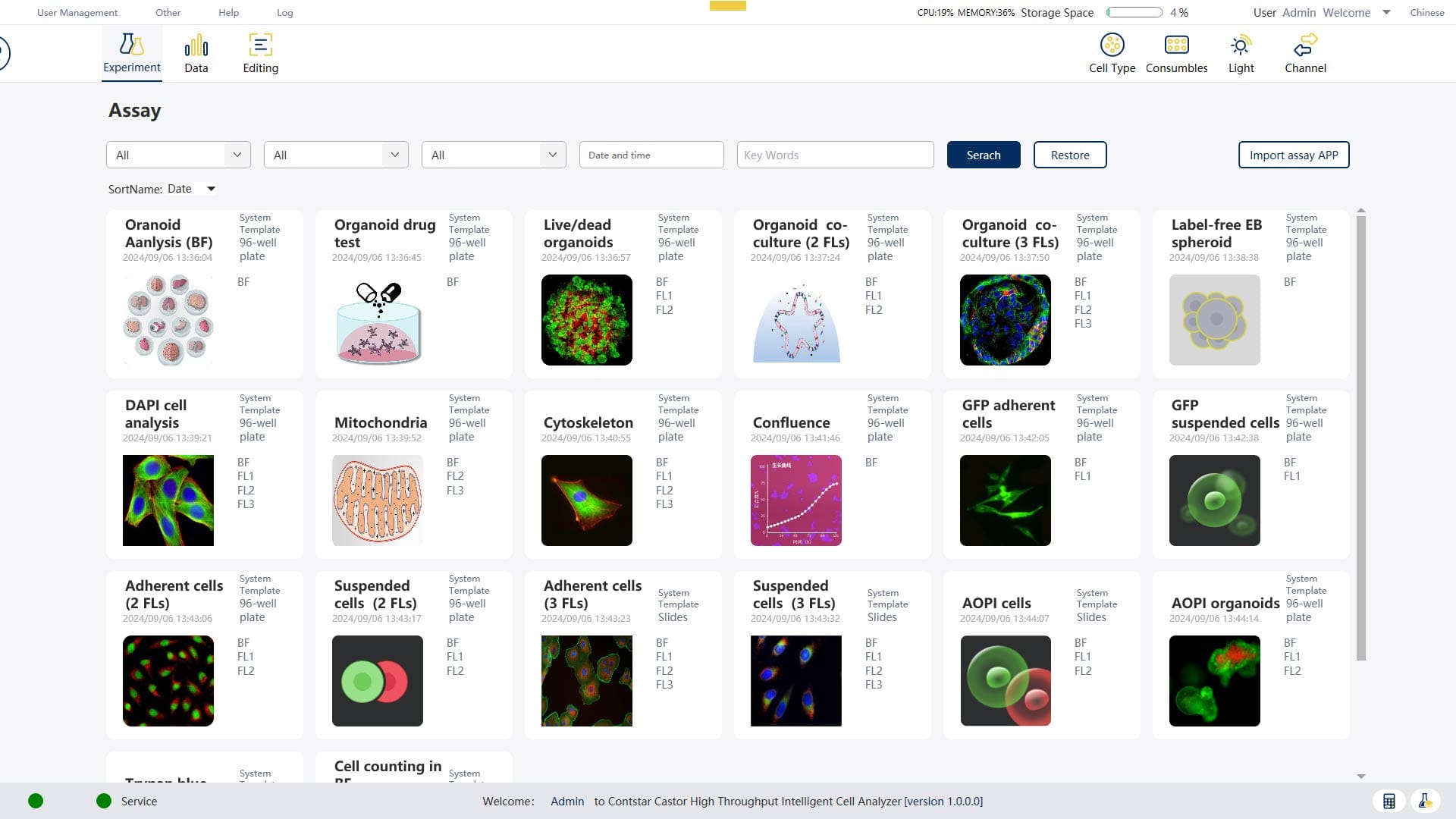
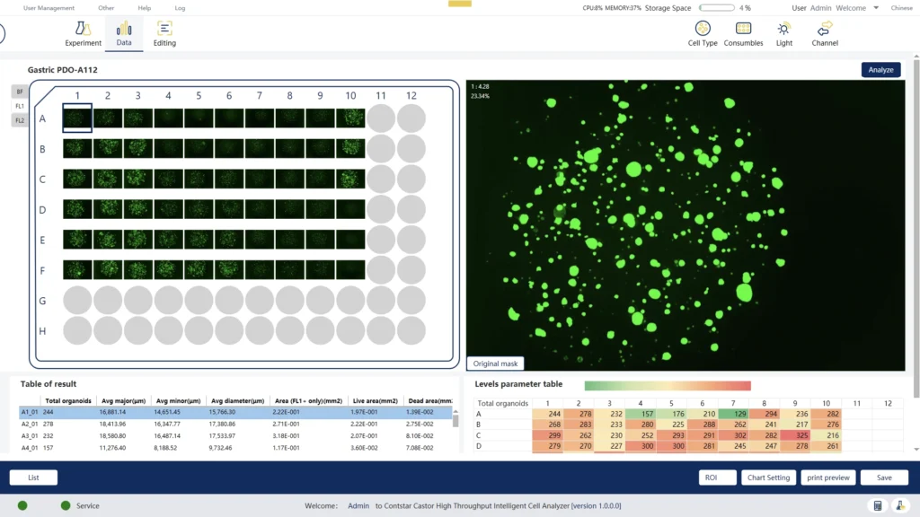
AI-Powered Cell Analysis
Leveraging deep learning-based image recognition to accurately segments and classifies target cells and organoids, enabling unbiased and deeper insights.

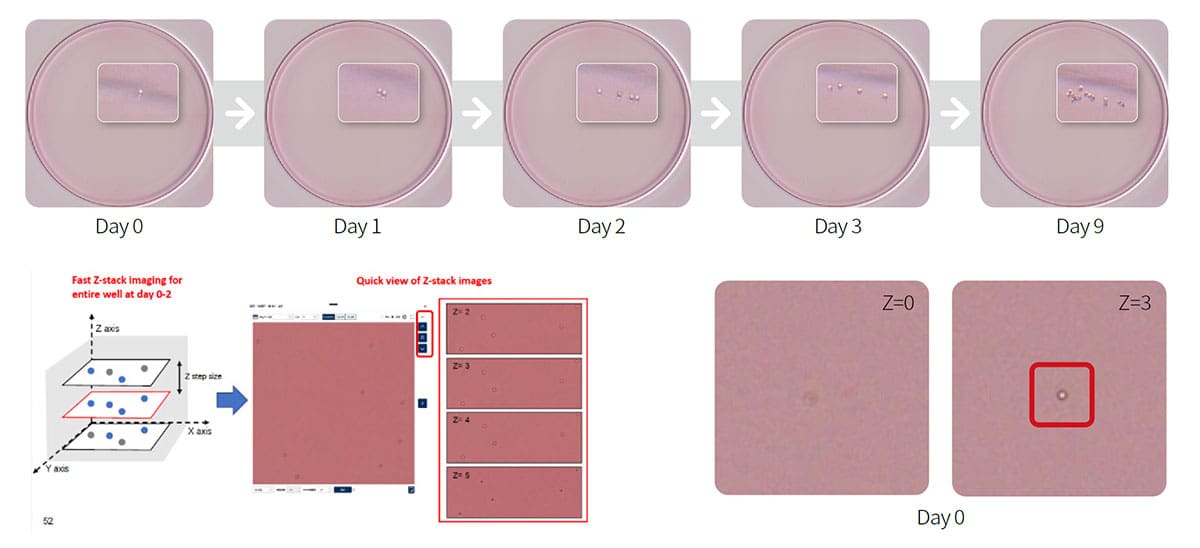
Ultrafast Z-Stack Imaging
Captures ten 2D images per well in just 4 minutes for a 96-well plate, significantly boosting your throughput and accelerate your discovery.

Rapid Autofocusing
Scans each well in milliseconds, adapting to variations in consumables to ensure pinpoint accuracy and consistent results.
Uniform Whole Well Imaging
Innovative concave meniscus illumination technology ensures uniform image quality across the entire well, enabling accurate identification and analysis of every single cell.

Compliance Ready
Offers robust data management and is fully compliant with FDA 21 CFR Part 11, ensuring the highest data integrity and reliability for your critical assays.

Automation Integration Ready
Seamlessly integrating with your existing automation solutions to dramatically increase the number of samples you can process.
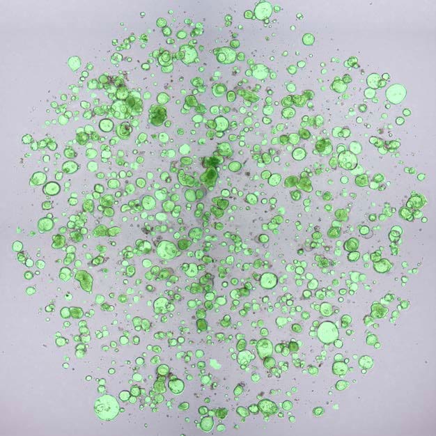
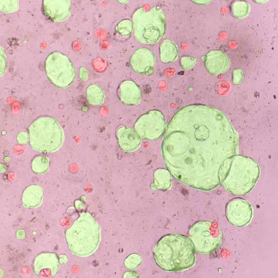
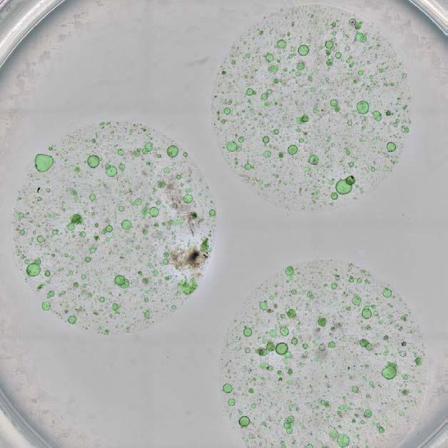
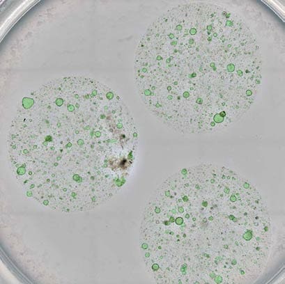
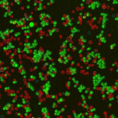
Organoid Analysis
- Organoid /Spheroid culture quality control
- Label-free organoid drug response test
- Organoid fluorescence viability analysis
- Cytotoxicity analysis for tumor organoid-based co-culture assay
Drug Discovery
- Hybridoma clone screening
- Single B cell clone counting
- GFP/RFP cell transfection
- Confluence analysis
- Cell proliferation, apoptosis, toxicity
Virus Titer Detection
- Plaque assay
- Virus titer detection by immunostaining (brown or blue spots)
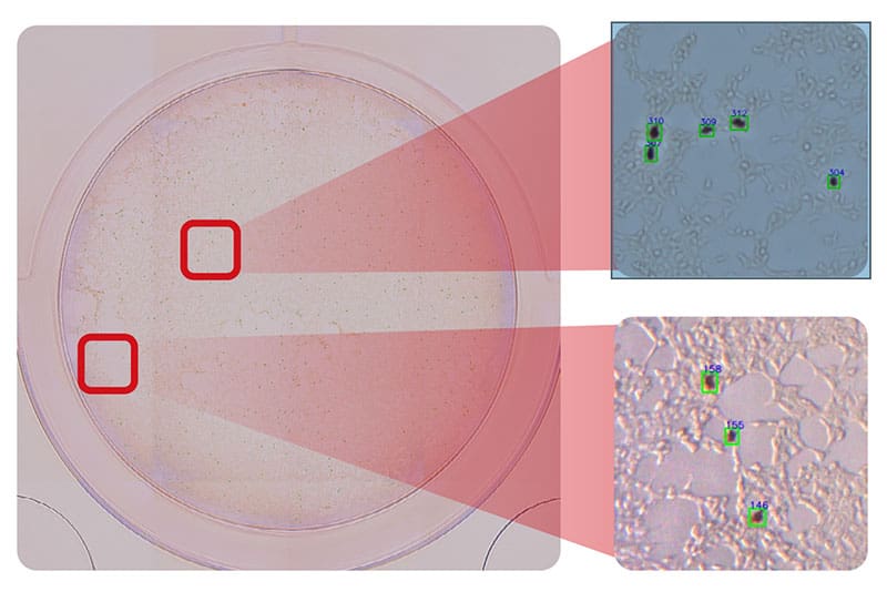
Cell Line Development
- Assurance of monoclonality
- GFP/RFP cell transfection
- Label free cell counting
- Viability analysis
- Confluence analysis