Countstar Spica M1

Opening the Door to Your Live Cell Analysis

A Tool For Every Lab

Incubator-Friendly for Optimal Cell Health

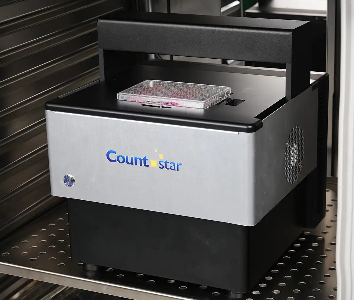
Fits directly inside any standard incubator, ensuring physiological conditions for reliable long-term imaging.

Monitor Experiments from Anywhere, Anytime

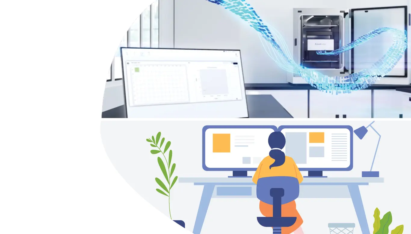
Stay connected to your ongoing experiments and track critical cellular events remotely, increasing flexibility and efficiency.

Ready-to-Go Assays

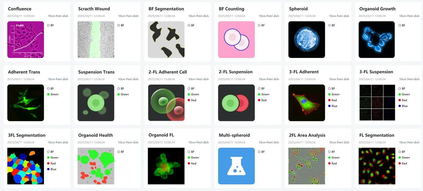
Start experiments faster with pre-set parameters for common applications and ensure consistent, reliable data for all users.

Seamless Imaging to Analysis, All in One

Integrated software streamlines your entire workflow, from acquisition to visualization. No need for multiple programs or extra costs.

Unparalleled Flexibility for Diverse Live Cell Assays

Unlock Deeper Insights from Complex Assays

Our integrated AI analysis tools efficiently process various datasets to uncover insights. They enable quick identification of key features and automated quantification, even in complex experiments that traditional methods struggle to analyze.

Support a Spectrum of Assays

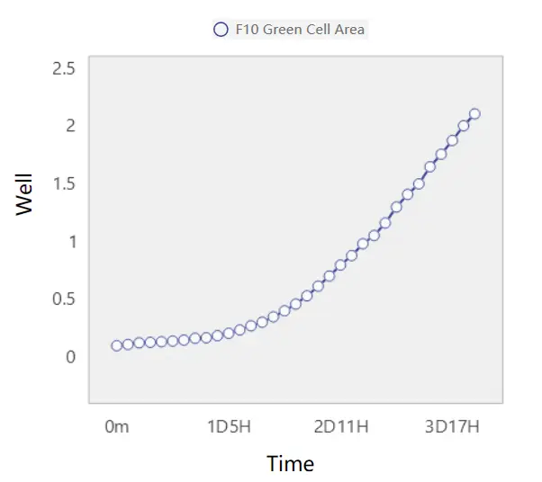
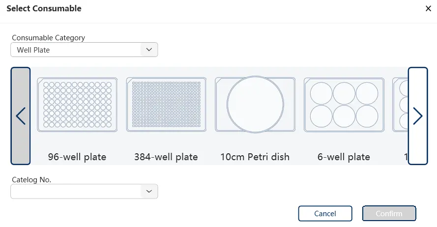
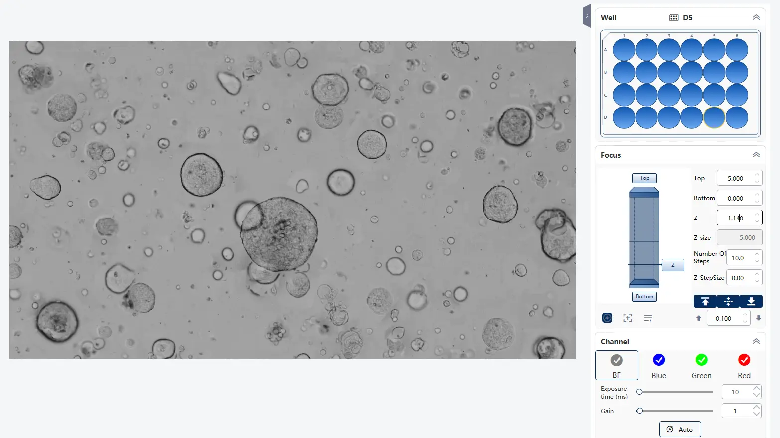
Image various cell types and markers simultaneously with multiple channels, observe at different scales with flexible magnification and utilize your preferred lab consumables.

Tailor Experiments to Your Specific Research

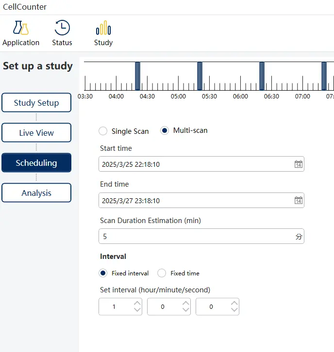
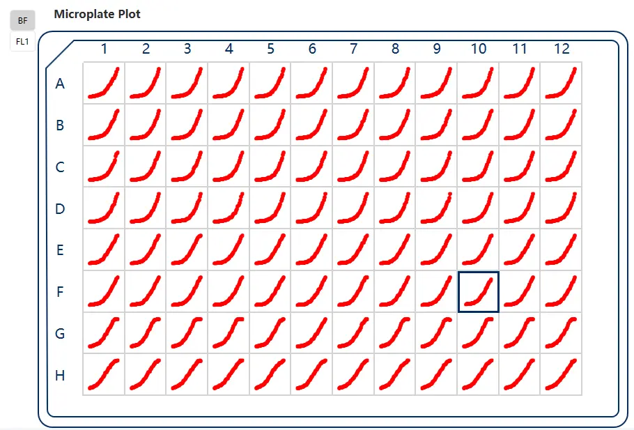
Customize imaging parameters and analysis metrics to meet the unique demands of your diverse assays, providing the exact data you need.

Capture Transient Events on Demand

Live Capture Mode allows easy observation and recording of dynamic, short-lived cellular processes in real time, providing immediate insights into critical biological events.

A Wide Range of Supported Assays

  • Label-Free Cell Counting
  • Reporter Genes
  • Proliferation
  • Scratch Wound
  • Organoid
  • Immune Cell Killing
  • Tumor Spheroid

Scratch Wound

Transfection

Tumor Spheroid

Organoid Growth

CameraHigh-sensitivity CMOS camera, 3200*2200, 7.1MP
Fluorescence channelEx:375/28,Em:430/30nm
Ex:470/20,Em:515/40nm
Ex:560/25,Em:630/70nm
Optical magnification and resolution7.5x: 0.6 µm/pixel
10x: 0.45 µm/pixel
15x: 0.30 µm/pixel
Field of view7.5x: 1.92*1.32mm
10x: 1.44*0.99mm
15x: 0.96*0.66mm
Focus ModeLaser focus and image focus
Imaging modeBright field, fluorescence; whole well scanning, fixed field of view/random field of view, Z-axis layer scanning
Supported consumables6-384 well plates, 35/60/100mm dishes, T25/T75 flasks, organ chips, microfluidic chips
Analysis AlgorithmAI image analysis and conventional image processing
Exported video formatAVI, MP4
Exported image formatJPEG,TIFF,BMP,PNG
Exported data formatXLSX, CSV
Dimensions300(L)*300(W)*337(H) mm
Computer recommendationsOS: Win10/11 Pro
CPU: i7-13700/ i7-14700 or higher
RAM: 64G or more
GPU: NVIDIA 1660 with 8G of VRAM or more
Storage: 8T or more

Request a Demo

We appreciate your interest in our instruments. Please provide your information below and we will get back to you shortly.

Disclaimer
This field is for validation purposes and should be left unchanged.

Request a Quote

We appreciate your interest in our instruments. Please provide your information below and we will get back to you shortly.

Disclaimer
This field is for validation purposes and should be left unchanged.