| Camera | High-sensitivity CMOS camera, 3200x2200, 7.1MP |
| Fluorescence channel | Ex:375/28,Em:430/30nm
Ex:470/20,Em:515/40nm
Ex:560/25,Em:630/70nm |
| Optical magnification and resolution | 7.5x: 0.6 µm/pixel
10x: 0.45 µm/pixel
15x: 0.30 µm/pixel |
| Field of view | 7.5x: 1.92x1.32mm
10x: 1.44x0.99mm
15x: 0.96x0.66mm |
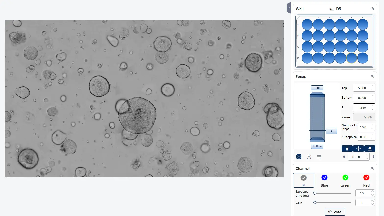
| Focus Mode | Laser focus and image-based focus |
| Imaging mode | Bright-field microscopy, Fluorescence microscopy, Z-axis layer scanning |
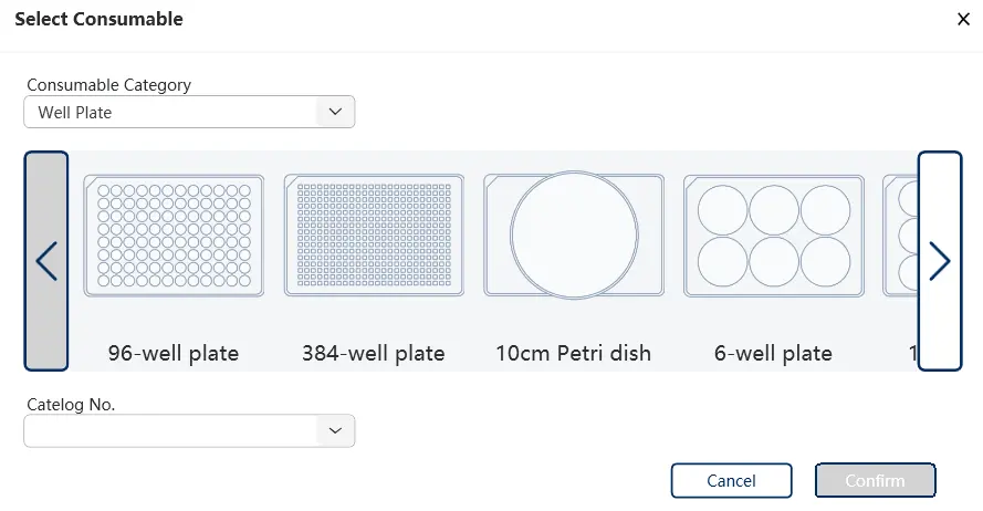
| Supported consumables | 6-384 well plates, 35/60/100mm dishes, T25/T75 flasks, organ-on-a-chip microfluidic chips |
| Analysis Algorithm | AI image analysis and conventional image processing |
| Exported video format | AVI, MP4 |
| Exported image format | JPEG, TIFF, BMP, PNG |
| Exported data format | XLSX, CSV |
| Dimensions | 300(L)x300(W)x337(H) mm |
| Computer recommendations | OS: Win10/11 Pro
CPU: i7-13700/ i7-14700 or higher
RAM: 128G or more
GPU: NVIDIA 4060 with 8G of VRAM or more
Storage: 4T SSD, 8T HDD or more |